Blackford Analysis
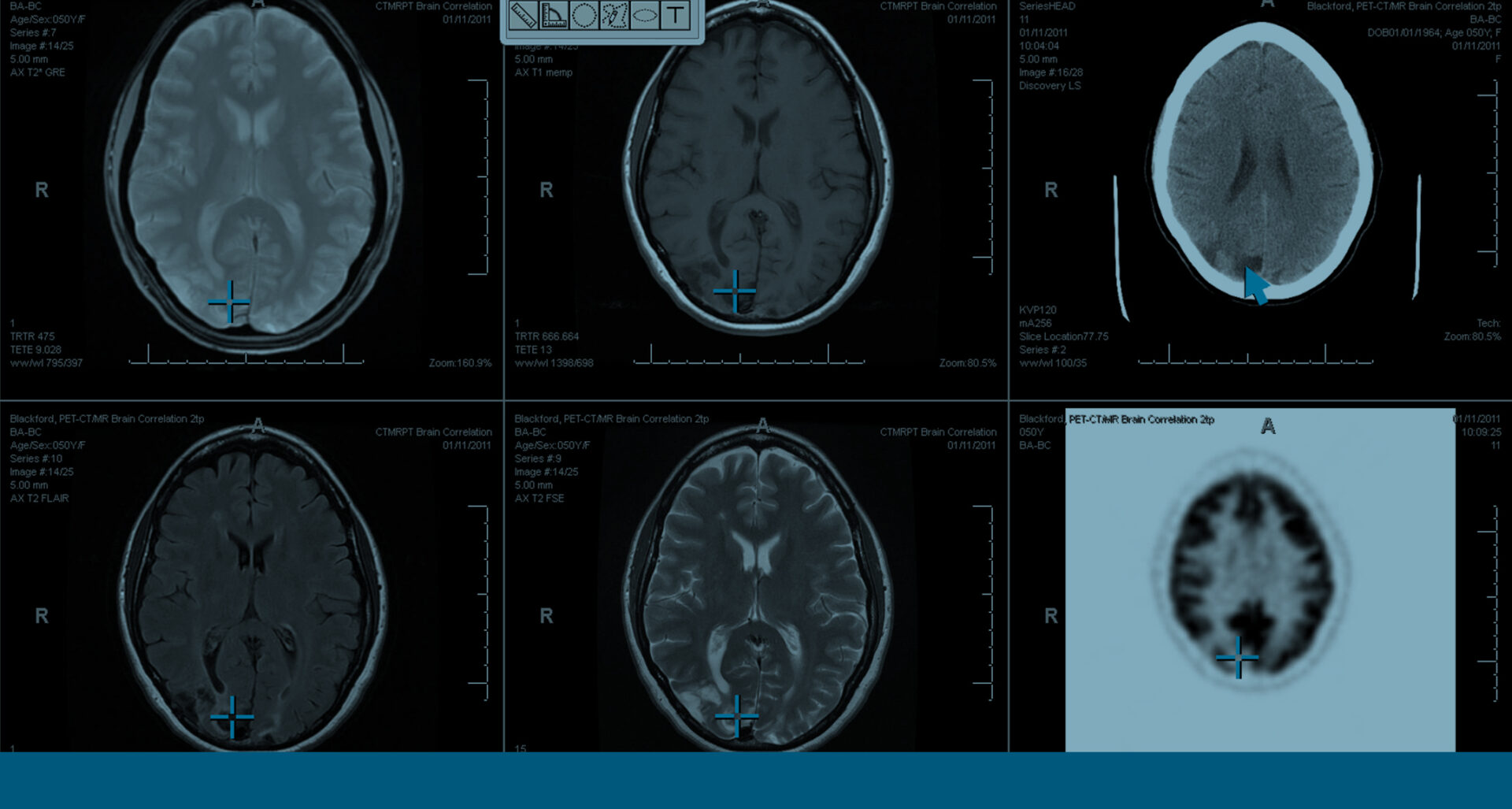
New enterprise image processing solutions have now become available to support the development of new clinical applications that have the ability to speed clinical workflow and facilitate the development of custom clinical applications that go beyond surgical and radiotherapeutic planning. The increasing emphasis on improving healthcare clinical quality and lowering costs are two of the many factors that have focused attention on the value of comparing a patient’s new imaging studies with all available relevant prior studies. The accepted clinical benefits of this are challenging radiologists to rapidly assimilate what can be a large amount of available information in order to maintain their productivity. These demands have created an opportunity for a new type of image registration. Automated registration automatically identifies and maps spatial locations across multiple imaging exams acquired at different times to create a new set of alignment data that enables any anatomy or pathology to be rapidly localized and observed across a series of current and previous study series.
Automated registration across the imaging enterprise The benefits and opportunities for automated registration may be most compelling within radiology, but the increasing conversation around enterprise imaging portends to the broader benefits of this technology. Because imaging is performed in many departments across the healthcare landscape opportunities to leverage automated registration may exist in departments such as neurology, oncology, and ophthalmology. Essentially, most clinical exams that benefit from imaging follow-up may benefit from automated registration, particularly when follow-up exams are performed on different imaging modalities and different locations. Another trend that is aligned with enterprise imaging is the ability to move pre-processing resources from a dedicated workstation to a centrally managed server whose IT resources can be centrally managed and shared across the organization. This enables a healthcare organization to more cost-effectively manage this resource and better integrate it with existing PACS environments or a centralized archive such as a Vendor Neutral Archive (VNA). This also eliminates isolated silos of post-processing capability that only work with certain dedicated systems, require specialized resources to manage and often provide limited access to the automated registration results. While standard PACS capability includes the ability to view current and prior exams on the same display, most PACS do not include the ability to process automated registration data. This is because specialized software and complex algorithms are required to enable the radiologist to realize the rapid feature identification and comparison capabilities that automated registration enables. Clinical tools such as these must be tightly and seamlessly integrated into the PACS workstation and the radiologist’s reading workflow to ensure that clinical productivity benefits are fully realized along with greater clinical confidence.
The language of automated registration As with most technologies, automated registration has a language all it’s own that warrants understanding. It is a technically complex process that entails alignment of 3D space, and identification of features in that space that may span imaging studies acquired at different times. Complicating matters is the fact that variations in patient positioning may cause anatomy or pathology to not precisely correlate with the same scanner location in each study. Two types of volumetric registration may be performed during the registration process: Rigid and Deformable. They each have their strengths and their unique applications. Following are the definition of key related terms that will strengthen your understanding of the image registration process and the unique characteristics of rigid and deformable registration.
Frame of Reference: Most medical images contain DICOM header information that describes where they exist in a discrete three-dimensional co-ordinate space known as a Frame of Reference:

- All images in a series exist in the same discrete three-dimensional co-ordinate space, identified by a Frame of Reference UID.
- Each image in a series also contains information that describes the image’s position and orientation within its Frame of Reference.


