Your strategic
AI platform partner
We provide tailored solutions to unlock the value of AI, drive efficiencies and improve patient outcomes.
Play Video
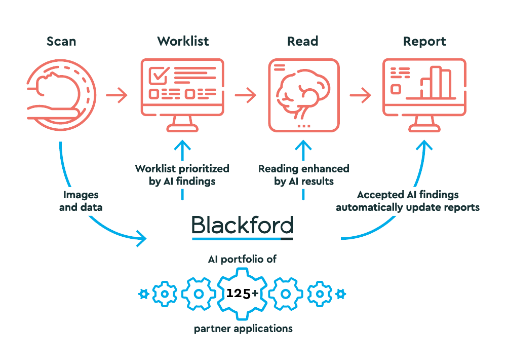
The Blackford Platform™ is our core focus and sits at the centre of our tailored AI offering.
Blackford Platform™ is purpose built for integration with your existing systems while simplifying integrations and the management of multiple disparate AI applications and algorithms, which ultimately reduces the load on imaging platforms. New AI applications from our market-leading portfolio, as well as in-house developed AI applications, can easily be added to the image-processing platform, reducing implementation time, costs, and long-term maintenance. Long term performance is monitored 24/7 by Blackford Dashboard™.

Core Subscription
Our Core Subscription provides access to a tried-and-tested core platform with the most comprehensive portfolio of Al solutions on the market. Also included is a range of features to tailor these Al solutions to your specific needs.

Professional services
In addition to the Core Subscription, our teams of clinical and technical experts can provide the following Professional Services to ensure the Al solutions best fit your requirements and deliver ongoing value.
AI Partner Portfolio
We currently have 125+ contracted AI applications across 50+ partner vendors covering 8 clinical and operational service lines.
Who we help

Non-Radiologist Clinicians
Clinicians, Neurologists, Oncologists, Orthopods and other referring Clinicians
Learn More >
Radiologist Clinicians
Radiologists, Radiology Directors, Technologists and other radiology staff
Learn More >
Administration
Senior hospital administrators including CIO, CMO, CMIO, CFO and legal counsel
Learn More >
Strategic Channel Partners
Establish a collaborative alliance for AI delivery in medical imaging.
Learn More >Latest Updates

Book a meeting
We’d welcome the opportunity to learn more about your AI needs and to explain how partnering with Blackford can drive efficiency and provide ongoing value.
Book a Meeting


